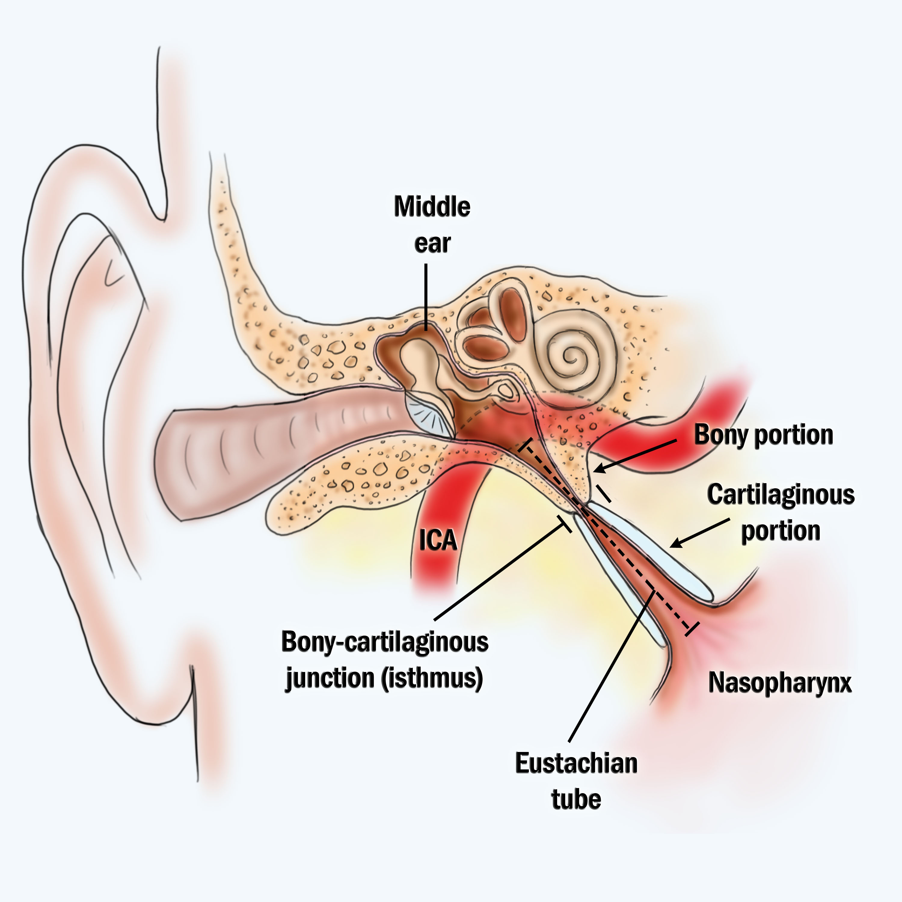

Vibrations of the vestibular membrane are transmitted through the malleus and incus to the stapes. The middle ear communicates with the mastoid air cells posteriorly and with the pharynx anteriorly through the Eustachian tube. Three auditory ossicles span the cavity between the tympanic membrane and an opening in the wall of the inner ear, the oval window. The middle ear, or tympanic cavity, is an air-filled space in the temporal bone that is lined by a mucosa.

Receives vibrations from stapes and transmits them to the oval windowĪbove: Structures of the outer, middle, and inner ear. Inferior to the oval window on the exterior of the cochlea, membrane-covered openingĬreates space for the fluid within the cochlea to move with the vibrations of the auditory ossiclesīetween incus and the oval window, stirrup-shaped bone, smallest bone in the human body Transfers vibrations from stapes to the cochlea Receives vibrations from the tympanic membrane and transmits them to incusīetween stapes and the cochlea, membrane-covered opening Receives vibrations from malleus and transmits them to stapesīetween the tympanic membrane and incus, hammer-shaped bone Middle auditory ossicles, anvil-shaped bone Passage between the middle ear and the nasopharynx (paired, one right and one left)Įqualizes the pressure of the middle ear with external air pressure (ear popping) The base of the stapes covers the oval window which allows sound waves to pass from the tympanic membrane, into the cochlea of the inner ear. The auditory ossicles inward from the tympanic membrane, are the malleus, incus, and stapes. The main structures of the middle ear are the auditory ossicles, Eustachian tube (also known as the pharyngotympanic tube), oval window and round window. Paired (right and left) membranes in the external auditory canal separating the outer ear from the middle earĮar drum sound causes vibration of the tympanic membrane vibrations of the tympanic membrane are transferred to the middle ear and then ultimately the inner ear for detection of sound Passageway carrying sound collected by the auricle to the middle ear Paired (right and left) skin-covered canal positioned within the external acoustic meatus (sometimes used synonymously with external acoustic meatus) Paired (right and left) canal in the bone of the skull, specifically the temporal boneĬreates passage within the skull for the external auditory canal Produce and secrete cerumen (ear wax) cerumen lubricates the external auditory canal and is antibacterial
#Auditory tube anatomy skin#
Located under the skin along the external auditory canal Paired (right and left) external ear structure composed of elastic cartilage and skinĬollecting sound and funneling it into the external auditory canal Elastic cartilage and the temporal bone form the support for the auricle. Additionally, ceruminous glands, specialized coiled, tubular, apocrine sweat glands, secret cerumen, a waxy secretion. Thin skin covering the external acoustic meatus possesses small dermal papillae and epithelial pegs to anchor the epidermis with the dermis. Numerous hair follicles with their accompanying sebaceous glands are also present. Skeletal muscle fibers of the auricular muscle have little, but variable, function in humans. The auricle or pinna (external ear) consists of a core of elastic cartilage covered by thin skin.


 0 kommentar(er)
